One defect map, multiple metrology tools
Combining CDSEM and high-magnification microscopy for smarter wafer defect detection
Wafer defect mapping becomes more effective when inspection results from different metrology tools are combined. By integrating CDSEM and high-magnification microscopy data, Nobleo enables a unified overview of defects on photonic wafers, supporting more efficient inspection workflows and targeted analysis of critical structures.
Combining metrology data for photonic wafer inspection
The validation of a Photonic Integrated Chip (PIC) requires a variety of different metrology tools. The metrology tools typically do both the imaging part as the processing part. Aggregating the results of different metrology tools and/or implementation of more complicated test flows might not be trivial because of the interface constraints imposed by the different tools.
Machine learning for aggregated wafer defect maps
We have developed software tools to create machine learning pipelines that process data from metrology tools and to aggregate the inspection results in a single defect map. A wafer quality engineer can visualize defects on function critical structures from different metrology tools in a single visual overview.
The tool feeds images from different metrology tools together with design information to a machine learning engine to create defect maps and scanning recipes. The feedback loop with scanning recipes allows quality engineers to create more advanced test flows: do a wafer scan and scan suspicious (or critical) areas with a CDSEM. Always, the output of the engine is a test report that contains a defect map which can be aggregated into a single GDS overlay.

Figure 1: An overview of the workflow used to combine image processing results from different metrology tools to create an aggregated defect map.
Combining CDSEM and high-magnification microscopy results
The workflow has been tested with CSEM in Switzerland. The idea is to combine high-magnification microscopy images with CDSEM data. The figure below shows a detail of a photonic wafer with aggregated defect maps from the two sensor modalities.


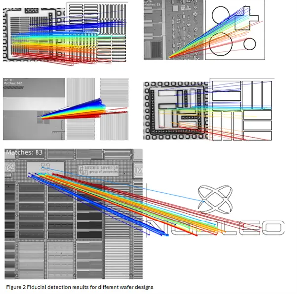
Figure 2: A set of rectangular areas has been defined for the different sensor modalities (left). The rectangular area has been scanned with a scanning microscope (middle). The picture on the right shows the defect map of the microscopy image on top of the CDSEM image. The cut-outs on the bottom right shows what the defect observed in the microscopy image looks like when imaged with a CD-SEM.
The software tool can be used to optimize test workflows to use the different pieces of equipment as efficiently as possible. The tool allows for efficient use of time consuming sensor modalities.
Detecting structures distorted by diffraction effects
The combination of CDSEM with high-magnification microscopy images can be used to train machine learning networks to “understand” microscopy images distorted by diffraction effects.
The network should be trained with a pairs of example images: one taken by the microscope and another taken with a CD-SEM. As long as the information required for the association is in the image, the machine learning algorithm will be able to model it.
There are more and more metrology applications where dimensions of critical structures are well below the diffraction limit. These design details are deformed because of diffraction artefacts in the microscopy images, which makes it difficult and time consuming to check inline with traditional methods.
A machine learning algorithm might be capable of modelling these deformed shapes as long as the measurements are sufficiently reproducible.
Particle detection (below diffraction limit)
State-of-the-art photonics often requires hybrid bonding. The presence of (smaller and smaller) particles introduces performance issues. Modelling of diffraction patterns with CDSEM and high-magnification microscopy can be used to measure particle sizes in the 10-100 nm range and it might even be feasible to classify these particles into different categories.


Figure 3 The figure on the left shows the defect map generated by the CD-SEM analysis. The image on the right shows a close-up of individual defects.
The combination of CDSEM with high-magnification microscopy data can be used to analyze which particles in the 10-100 nm range are still visible in the high-magnification microscopy images to understand the limits of what can be done with a microscopy set-up. As a second step, it might be feasible to train a machine learning network to predict the type and size of these particles.
High-magnification microscopy could be a suitable technology to speed-up inline testing and thereby increase the throughput for industrial test applications.
Metrology features
The combination of CDSEM and high-magnification microscopy images can also be used to monitor process variations.


Figure 4 The figure above shows a set of overlay markers that are used in wafer manufacturing. These markers are used typically to measure process parameters.
Training a network on the combination of diffraction deformed microscopy images and a CDSEM ground truth might be used to squeeze out a bit of additional alignment accuracy.
What could undetected wafer defects be costing you? Missed defects can lead to costly rework, delays and reduced yield. Fill in our contact form to explore how Nobleo can help you tackle these challenges.